Decoding the Precision Strike: How Killer T Cells Eliminate Cancer Cells
Introduction
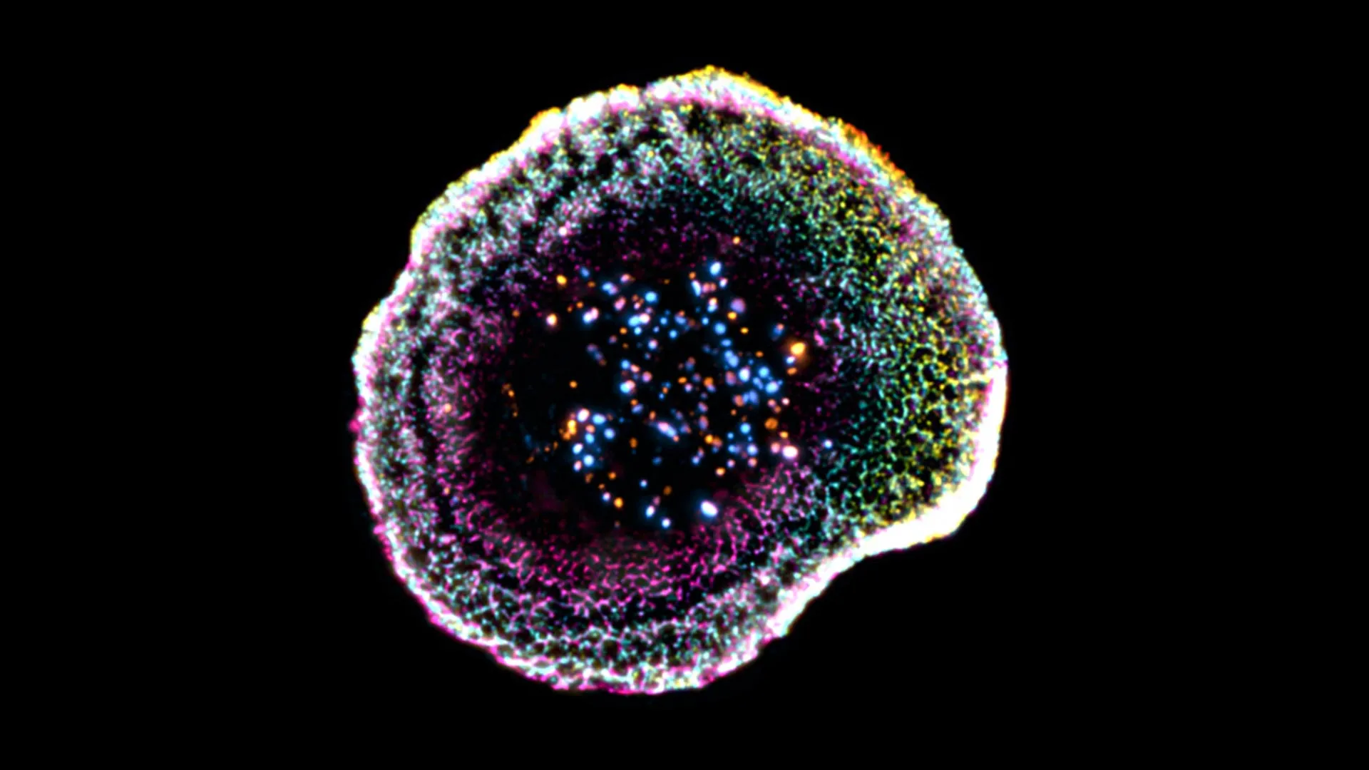
Imagine a microscopic assassin so precise that it can destroy a single diseased cell without disturbing its healthy neighbors. That's exactly what your body's killer T cells do every day. Thanks to a breakthrough 3D imaging technique, scientists have finally captured the intricate choreography behind this lethal efficiency. This guide breaks down—step by step—the molecular ballet that allows killer T cells to recognize, lock onto, and dismantle cancer cells with stunning accuracy. Whether you're a biology enthusiast, a student, or a curious reader, you'll gain a front-row seat to one of the immune system's most elegant performances.
What You Need
- Basic knowledge of cell biology – Familiarity with concepts like cell membranes, proteins, and DNA will help you follow the mechanics.
- An understanding of the immune system – Know the roles of T cells, B cells, and antigen presentation. If not, a quick primer on MHC molecules is recommended.
- A curious mind – No lab equipment required—just the willingness to visualize molecular events on a nanometer scale.
- Optional: Access to 3D molecular models – Online resources like the Protein Data Bank can bring these steps to life in interactive 3D.
Step-by-Step Instructions
Step 1: Patrol and Recognize the Invader
Killer T cells constantly roam the body, scanning for trouble. Their surface is covered with T cell receptors (TCRs), which act like molecular barcode scanners. Every healthy cell displays pieces of its internal proteins on its surface using MHC class I molecules. When a cell becomes cancerous, it presents abnormal peptides—mutated proteins. The TCR binds specifically to these cancer peptide–MHC complexes. This initial docking is weak and reversible, but it's the crucial first handshake that sets everything in motion.
Step 2: Form the Immunological Synapse
Once the TCR locks onto its target, the T cell doesn't just attack haphazardly. Instead, it reorganizes its surface molecules into a highly organized structure called the immunological synapse. Imagine a bullseye: the center, or central supramolecular activation cluster (cSMAC), is where TCRs cluster. Around it, adhesion molecules like LFA-1 bind to ICAM-1 on the cancer cell, forming a tight ring. This segregated arrangement ensures that the lethal payload will be delivered exactly at the point of contact, minimizing collateral damage to bystander cells.
Step 3: Polarize the Killing Machinery
Inside the T cell, a dramatic reorganization occurs. The cell’s microtubule-organizing center (MTOC) moves toward the synapse, towing a trail of vesicles filled with deadly cargo. These vesicles, called cytotoxic granules, contain two key weapons: perforin (which punches holes in the cancer cell membrane) and granzymes (serine proteases that trigger apoptosis). The T cell essentially aims its internal artillery at the exact point of contact, ready to fire.
Step 4: Release the Cytotoxic Granules
Upon receiving the final activation signals, the T cell fuses its granules with the plasma membrane at the synaptic cleft. Perforin molecules insert themselves into the cancer cell membrane, polymerizing to form pores. Through these pores, granzymes enter the target cell. The entire release is tightly controlled; multiple regulatory proteins ensure that the granules dock precisely at the contact zone before exocytosis occurs. This step is the reason the attack is so precise—no stray granules leak into the surrounding environment.
Step 5: Induce Programmed Cell Death
Inside the cancer cell, granzymes get to work. They cleave and activate caspases, the executioner enzymes of apoptosis. Additionally, granzyme B directly cuts proteins that maintain the cell’s survival pathways. The cancer cell begins to shrink, its DNA fragments, and its membrane blebs. Within minutes, the cell dies quietly and is promptly cleared by macrophages—no inflammation, no mess. The T cell, meanwhile, survives intact and can move on to find the next target.
Step 6: Detach and Recycle
After delivering the lethal blow, the killer T cell needs to disengage. Adhesion molecules like LFA-1 loosen their grip, and the immunological synapse disassembles. The T cell’s MTOC moves back to its usual position. The cell now recycles its surface receptors and prepares for another round of killing. Some T cells can kill multiple cancer cells in sequence, making them highly efficient serial killers.
Tips for Understanding and Application
- Visualize the nanoscale choreography – Watch animations of the immunological synapse online. The 3D view that scientists captured reveals how the molecules arrange themselves like a perfectly orchestrated dance.
- Connect to immunotherapy – Many modern cancer treatments, such as checkpoint inhibitors and CAR-T cell therapy, enhance or mimic these steps. Understanding the natural process helps you grasp how these therapies work.
- Appreciate the precision – The reason T cells don't harm healthy neighbors is the tight spatial control of granule release. Remember, the synapse acts as a molecular funnel, concentrating the attack.
- Think about failures – Tumors often evade this process by downregulating MHC or secreting suppressive signals. This knowledge is key to developing new strategies to overcome resistance.
- Explore further – Search for “killer T cell 3D imaging” to see the actual experimental data. The first-ever high-resolution view of this process was a landmark in immunology.