A Step-by-Step Guide to Reversing Diabetes in Mice Using Lab-Grown Insulin Cells
Introduction
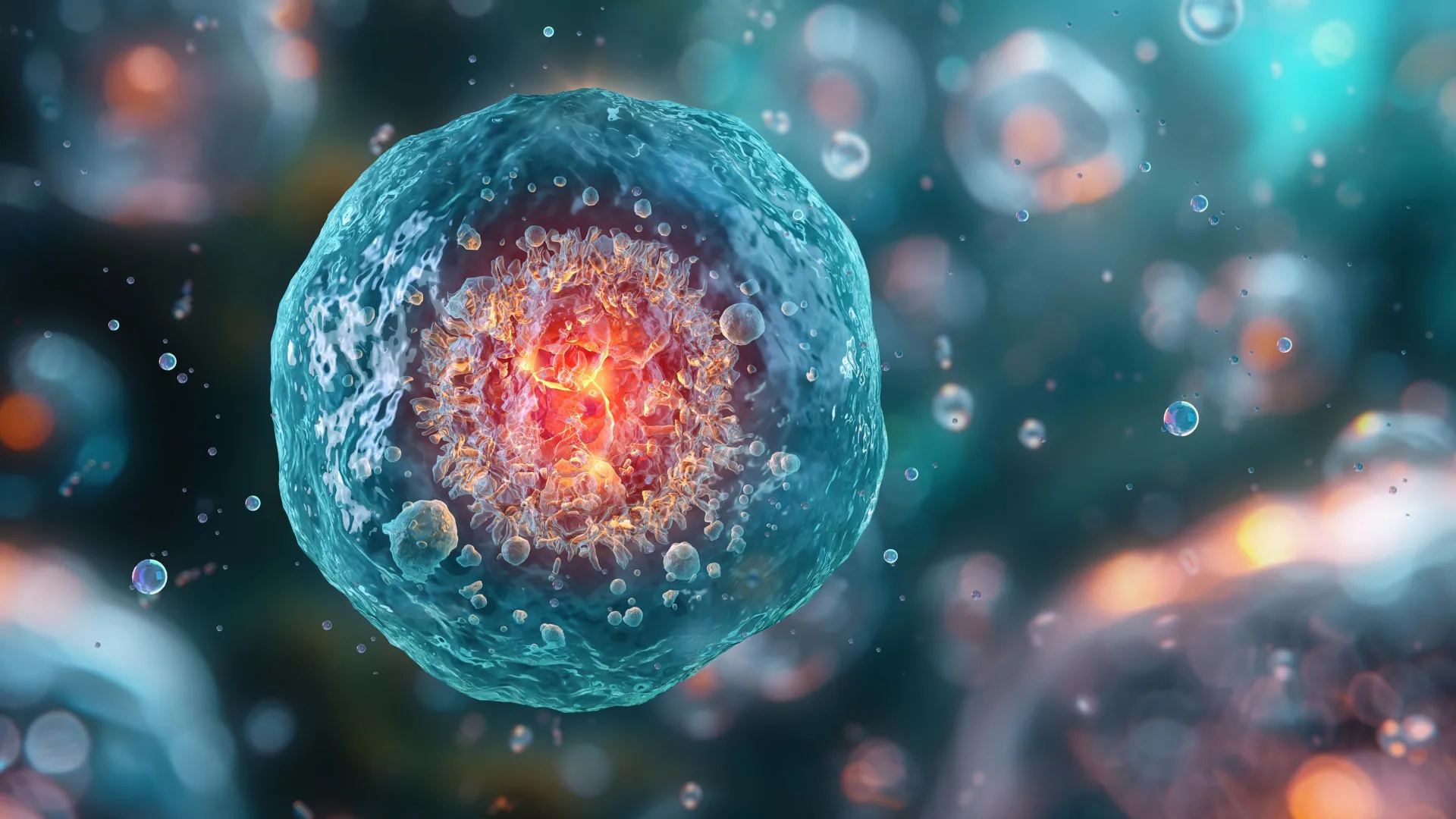
Type 1 diabetes affects millions worldwide, but a groundbreaking study by Swedish scientists has brought us closer to a functional cure. By creating insulin-producing cells from human stem cells, researchers successfully restored blood sugar control in diabetic mice. This guide breaks down the key steps involved in this revolutionary process, from cell creation to transplantation. Whether you're a student, researcher, or curious reader, follow along to understand how lab-grown cells could one day transform diabetes treatment.
What You Need
Before diving into the steps, gather the essential materials and prerequisites. Note that this is a specialized laboratory process requiring strict sterile conditions and ethical approvals.
- Human pluripotent stem cells (e.g., induced pluripotent stem cells or embryonic stem cells) – ethically sourced and approved for research.
- Differentiation media and growth factors – specific cocktails to guide stem cells toward pancreatic beta cell lineage.
- Culture dishes and incubators – with controlled temperature, humidity, and CO2 levels (37°C, 5% CO2).
- Glucose stimulation test kit – to measure cell response to varying glucose concentrations.
- Immune-deficient diabetic mice – e.g., NOD-SCID mice with chemically induced diabetes (streptozotocin model).
- Transplantation tools – sterile syringes, surgical microscope, and encapsulation materials (e.g., alginate microcapsules) if needed.
- Blood glucose monitoring system – for regular measurement before and after transplantation.
- Ethical approval – from institutional animal care and use committee (IACUC) and stem cell research oversight.
Step-by-Step Procedure
Step 1: Prepare and culture human pluripotent stem cells
Begin with a reliable source of human pluripotent stem cells (hPSCs). Thaw a vial of cryopreserved cells and plate them on a feeder layer (e.g., mouse embryonic fibroblasts) or in a feeder-free system using appropriate matrix (like Matrigel). Maintain the cells in stem cell medium (e.g., mTeSR1) and passage them when they reach 70–80% confluence. Monitor daily for spontaneous differentiation and remove any colonies that deviate from the typical morphology. Keep the cells in a humidified incubator at 37°C with 5% CO2. This step ensures a homogeneous starting population for differentiation.
Step 2: Differentiate stem cells into insulin-producing cells
Once you have a healthy, undifferentiated culture, initiate the differentiation protocol. The Swedish team used a multi-stage approach that mimics embryonic pancreatic development. Over 20–30 days, apply a series of growth factors and small molecules in a specific temporal order:
- Definitive endoderm induction (days 1–4): Add Activin A and Wnt3a to drive cells toward endoderm.
- Primitive gut tube formation (days 4–7): Switch to FGF7 and retinoic acid to direct cells toward pancreatic fate.
- Posterior foregut specification (days 7–14): Use retinoic acid, FGF10, and Noggin to create pancreatic progenitor cells.
- Pancreatic endoderm and endocrine induction (days 14–20): Supplement with EGF, nicotinamide, and T3 to generate insulin-producing beta-like cells.
- Maturation (days 20–30): Culture in a maturation medium containing high glucose, Sigma, and other additives to enhance glucose responsiveness.
At each stage, confirm successful differentiation using markers (e.g., PDX1, NKX6.1, insulin) via immunostaining or qPCR.
Step 3: Assess glucose-stimulated insulin secretion (GSIS)
Before using the cells in animals, verify they respond to glucose changes – a key feature of healthy beta cells. Harvest a batch of differentiated cells and incubate them first in low-glucose medium (2 mM) for one hour, then in high-glucose medium (20 mM) for another hour. Collect the supernatant at each step and measure insulin concentration using ELISA. A robust response shows a significant increase (at least 3–5 fold) in insulin release upon high glucose exposure. For the Swedish study, the lab-grown cells showed strong glucose responsiveness, making them suitable for transplantation.
Step 4: Prepare diabetic mice for transplantation
Obtain immune-deficient mice (e.g., NOD-SCID) to prevent rejection of the human cells. Induce diabetes by a single injection of streptozotocin (150–200 mg/kg intraperitoneally) after fasting. Confirm diabetes by measuring blood glucose levels > 300 mg/dL for two consecutive days. Maintain the mice under sterile conditions (IVC cages) and monitor body weight daily. For long-term experiments, provide supplemental insulin (0.1–0.2 U/day) to prevent severe weight loss until transplantation.
Step 5: Transplant the lab-grown insulin cells into diabetic mice
Anesthetize a diabetic mouse using isoflurane. Prepare the cells: detach them from culture dishes with TrypLE, wash, and resuspend in a small volume (10–20 µL per mouse) of cold saline. Optionally encapsulate the cells in alginate microcapsules to reduce immune response and facilitate retrieval. Using a surgical microscope, perform a renal subcapsular transplantation (common site for minimal invasiveness) or an intraportal injection into the liver. Make a small incision in the left flank, expose the kidney, and inject the cell suspension under the capsule using a fine needle. Close the incision with absorbable sutures. Allow the mouse to recover on a heat pad.
Step 6: Monitor blood glucose normalization
Post-transplantation, measure non-fasting blood glucose daily using a tail vein glucometer. The Swedish researchers observed that blood glucose levels dropped gradually and reached normal range (< 200 mg/dL) within 2–3 weeks. Continue monitoring for at least four weeks to confirm sustained control. Optionally, perform a glucose tolerance test (GTT) at the endpoint: fast mice for 6 hours, inject glucose (2 g/kg i.p.), and measure glucose at 0, 15, 30, 60, and 120 minutes. Successful reversal shows improved glucose clearance compared to diabetic controls. Confirm the presence of human C-peptide (co-secreted with insulin) in mouse serum as evidence that the transplanted cells are producing insulin.
Step 7: Evaluate long-term outcomes and safety
At the end of the study (typically 8–12 weeks), euthanize the mice humanely and collect the transplanted tissue for histological analysis. Stain for insulin, glucagon, and other endocrine markers to confirm cell identity and engraftment. Check for any tumor formation (teratomas) due to residual undifferentiated stem cells – the Swedish team reported none, indicating a well-differentiated population. Finally, share your findings in a peer-reviewed journal, following ARRIVE guidelines for animal research.
Tips for Success
- Optimize differentiation efficiency: Small variations in growth factor timing or concentration can drastically affect yield. Use a serum-free and defined medium to improve reproducibility.
- Purify beta-like cells: Before transplantation, consider sorting cells using surface markers (e.g., CD49a, CD200) to enrich insulin-positive cells and reduce heterogeneity.
- Use immune-compromised mice: Human cells are xenografts; standard mice will reject them. NOD-SCID or NSG strains are essential.
- Monitor for hypoglycemia: Overproduction of insulin can cause dangerous lows. Start with a moderate cell dose (1–2 million cells per mouse) and titrate if needed.
- Document everything: Keep a detailed lab book with dates, media compositions, and observations. This will help troubleshoot and replicate results.
- Collaborate with experts: If you’re new to stem cell differentiation or rodent surgery, seek mentorship from an experienced lab – the protocol is complex and requires precision.
Note: This guide is based on publicly available research from the study published by Swedish scientists. Always follow your institutional ethics guidelines and regulatory requirements before attempting any animal or stem cell experiments.